What Is PACS?
A Picture Archiving and Communication System (PACS) is a medical imaging technology used to store, retrieve, distribute, and display images from various modalities (X-ray, CT, MRI, ultrasound, nuclear medicine, fluoroscopy). PACS replaced traditional film-based radiology with digital workflows, fundamentally transforming how medical images are acquired, stored, reviewed, and shared.
The four major components of any PACS are:
- Image acquisition modalities — the radiology devices that generate images (X-ray, CT, MRI, ultrasound, PET, mammography)
- A secure network — the digital infrastructure connecting modalities to storage and workstations
- A storage archive — short-term and long-term storage for medical images
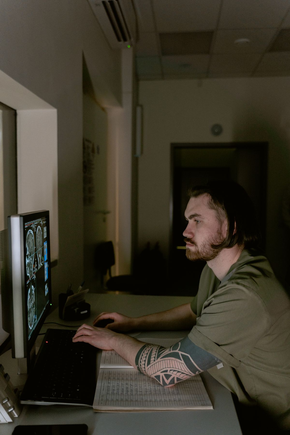
- Integrated display workstations — diagnostic and clinical review stations for radiologists and physicians

---
Understanding DICOM: The Language of PACS
What Is DICOM?
DICOM (Digital Imaging and Communications in Medicine) is the international standard (ISO 12052) that defines how medical images and associated metadata are formatted, transmitted, and stored. Every modern radiology device — regardless of manufacturer — speaks DICOM. Without DICOM, a GE CT scanner could not send images to a Siemens PACS, and a Philips MRI could not be read on an Agfa workstation.
A DICOM file contains two things bundled together: the pixel data (the actual image) and a header containing patient demographics, study information, acquisition parameters, and device identification. This integration ensures images are never separated from their identifying information — a critical patient safety feature.
Key DICOM Services for Device Connectivity
| DICOM Service | Function | Direction |
|---|---|---|
| C-STORE | Transfers an image from modality to PACS | Modality → PACS |
| C-FIND | Queries for studies/series/images | Workstation → PACS |
| C-MOVE | Requests transfer of images to a destination | Workstation → PACS |
| C-GET | Pulls images directly to the requesting system | Workstation → PACS |
| Modality Worklist (MWL) | Retrieves patient/order list to modality | PACS → Modality |
| Modality Performed Procedure Step (MPPS) | Reports exam status back to PACS | Modality → PACS |
| Storage Commitment | Confirms PACS has safely stored images | PACS → Modality |
How DICOM Association Works
When a modality sends images to PACS, it establishes a DICOM Association — a TCP/IP connection where both systems negotiate which services and data types they support. The process follows these steps:
- Association Request (A-ASSOCIATE-RQ): The modality sends a connection request specifying its AE Title, the PACS AE Title, and the services it wants to use
- Association Accept (A-ASSOCIATE-AC): PACS responds, accepting or rejecting each proposed service
- Data Transfer: Images are sent as C-STORE commands, each containing a DICOM dataset
- Association Release (A-RELEASE-RQ/RP): The connection is gracefully closed

---
Connecting Radiology Devices to PACS
Step 1: Network Configuration
Every radiology device and PACS component must be on a network that can communicate. In most hospitals, a dedicated imaging VLAN separates radiology traffic from general hospital traffic to ensure bandwidth and security.
Required network parameters for each modality:
- IP Address: Static IP on the imaging network (never DHCP for medical devices)
- Subnet Mask and Gateway: Matching the imaging VLAN configuration
- AE Title: A unique Application Entity title identifying the device (e.g.,
CT_SCANNER_01) - Port Number: Typically 104 (standard DICOM) or 11112 (DICOM TLS secure)
- PACS AE Title and IP: The destination PACS address configured on the modality
Step 2: Modality Worklist Configuration
The Modality Worklist (MWL) is the first integration point between a radiology device and PACS/HIS. When a patient is registered in the Hospital Information System (HIS) and an order is placed in the Radiology Information System (RIS), this information becomes available as a worklist entry.
How it works in practice:
- A technologist walks up to the CT scanner and selects "Worklist" on the console
- The CT scanner queries the MWL server (usually RIS or PACS) using C-FIND
- The worklist displays all scheduled exams for today, filtered by modality type
- The technologist selects the correct patient and exam
- Patient demographics and exam parameters are automatically loaded — no manual entry
- After the scan, images are sent to PACS with the correct patient and study context
This workflow eliminates transcription errors — a critical safety feature when dealing with patient identities.
Step 3: Image Transfer Configuration (C-STORE)
After images are acquired, the modality sends them to PACS using C-STORE. The configuration on each modality includes:
- Destination AE Title: The PACS AE Title (e.g.,
PACS_MAIN) - Destination IP Address: The PACS server IP
- Destination Port: Usually 104 or 11112
- Transfer Syntaxes: The encoding format (Implicit VR Little Endian, Explicit VR Little Endian, JPEG Lossless, etc.)
Transfer Syntax negotiation is a common source of connectivity issues. The modality and PACS must agree on how image data is encoded. Most modern systems support:
- Implicit VR Little Endian (1.2.840.10008.1.2): Default, universally supported
- Explicit VR Little Endian (1.2.840.10008.1.2.1): Preferred for most images
- JPEG Lossless (1.2.840.10008.1.2.4.70): For compressed images, reduces bandwidth
- JPEG 2000 Lossless (1.2.840.10008.1.2.4.90): Advanced compression for large studies
Step 4: Storage Commitment
After sending images, the modality requests Storage Commitment — a formal confirmation that PACS has received and stored the images. This is a legal and safety requirement: a technologist should not delete images from the modality until PACS confirms safe storage.
The process:
- Modality sends a Storage Commitment Request (N-ACTION) listing the images
- PACS verifies all images are in its archive
- PACS sends a Storage Commitment Result (N-EVENT-REPORT) confirming success or failure
- Only after confirmation can the modality safely purge local images
---
Connecting Specific Radiology Modalities
X-Ray (Digital Radiography and Fluoroscopy)
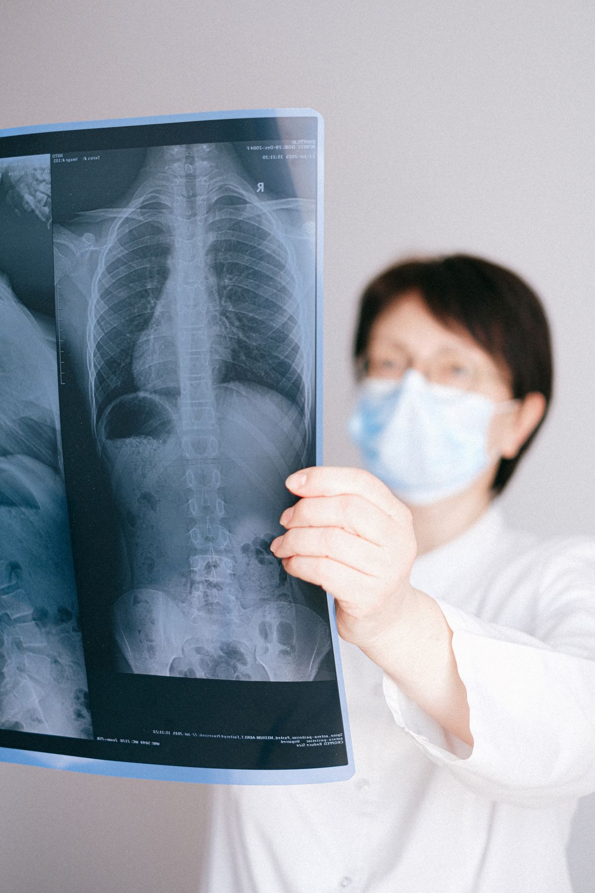
Digital X-ray systems (DR) use flat-panel detectors that produce DICOM images directly. Connection to PACS is straightforward:
- CR (Computed Radiography): Cassette-based systems where plates are read by a scanner, then images are sent to PACS
- DR (Digital Radiography): Direct digital capture, images appear within seconds and are sent to PACS automatically
- Fluoroscopy: Real-time X-ray imaging; studies may include both cine loops (video) and spot images, all sent as DICOM
Common configuration issue: X-ray images are often large (35×43 cm plates at high resolution). Ensure the network can handle the bandwidth — a single chest X-ray can be 10-30 MB.
CT (Computed Tomography)
CT scanners produce large studies — a single exam may contain 1,000-5,000 slices. Key connectivity considerations:
- Multi-slice transfer: Images are sent as they are reconstructed, not all at once
- Series organization: A CT study has multiple series (e.g., scout, axial, coronal, sagittal reconstructions)
- MPPS reporting: CT reports exam progress to PACS, enabling real-time workflow tracking
- Dose reporting: Modern CT scanners include DICOM Radiation Dose Structured Reports (RDSR)
Bandwidth tip: A CT study can be 500 MB - 2 GB. Ensure the modality-to-PACS link is gigabit or higher.
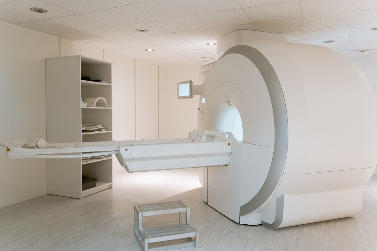
MRI (Magnetic Resonance Imaging)
MRI studies are complex with multiple sequences, contrasts, and reconstructions:
- Multiple series per study: T1, T2, FLAIR, DWI, etc. — each is a separate DICOM series
- 3D acquisitions: Large volume datasets requiring significant storage
- Enhanced MR objects: Some scanners use Enhanced DICOM objects that pack multiple series into one multiframe file
Configuration note: MRI transfer syntaxes often include JPEG Lossless or JPEG 2000 to compress large datasets. Verify PACS supports the specific transfer syntax the MRI uses.
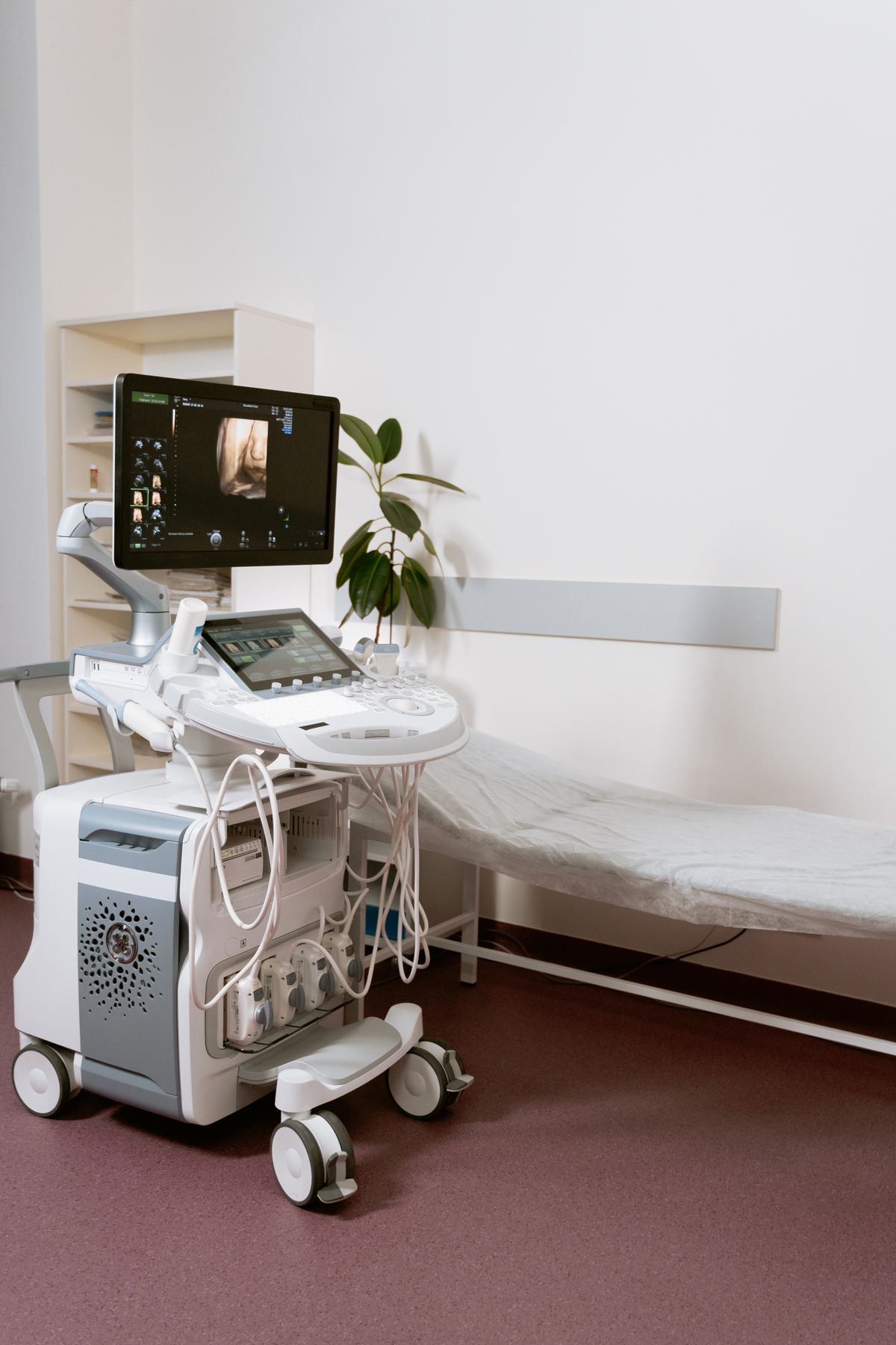
Ultrasound
Ultrasound connectivity has unique challenges:
- DICOM encapsulation: Ultrasound images may include measurements, annotations, and calculations embedded in DICOM structured reports
- Cine loops: Ultrasound captures video clips as multiframe DICOM objects
- SR (Structured Reports): Measurements like biometry, cardiac calculations, and Doppler values are sent as DICOM SR objects
- Connectivity issue: Some older ultrasound machines use proprietary formats. A DICOM gateway or converter may be needed
Mammography
Mammography produces the largest single images in radiology:
- Image size: A single mammogram can be 50-100 MB (high resolution is mandatory for detecting microcalcifications)
- DICOM Mammography IOD: Uses specific DICOM objects with mammography-specific attributes (view position, compression force, breast thickness)
- Tomosynthesis (3D mammography): Produces 3D volume datasets — a single study can be 1-3 GB
- Network requirement: Dedicated high-bandwidth connection; consider compression only with lossless transfer syntaxes
Nuclear Medicine and PET
PET and nuclear medicine have specific DICOM requirements:
- DICOM PET IOD: Includes radiopharmaceutical information, uptake time, decay correction
- PET-CT fusion: Combined studies where PET and CT data must be correlated; PACS must support fused image display
- Non-image data: Dose calibration factors and quality control data may be sent as DICOM SR
---
PACS Network Architecture
Typical Hospital PACS Network Design
A well-designed PACS network follows a layered architecture:
Layer 1 — Acquisition Network:
- Dedicated VLAN for all modalities
- Gigabit switches with QoS prioritizing DICOM traffic
- Modalities connected via Cat6 copper or fiber
Layer 2 — Core Network:
- 10-gigabit backbone connecting acquisition to PACS servers
- Redundant paths for fault tolerance
- Firewall between imaging network and hospital network
Layer 3 — Distribution Network:
- Gigabit to diagnostic workstations
- 100 Mbps minimum to clinical review stations
- Wireless access for portable device review (tablet PACS viewers)
Layer 4 — External Network:
- Secure VPN for teleradiology and remote reading
- HL7/DICOM gateway for external image exchange
- Cloud sync for disaster recovery replication
Bandwidth Planning
| Modality | Average Study Size | Studies/Day | Daily Bandwidth |
|---|---|---|---|
| X-ray (DR) | 20 MB | 150 | 3 GB |
| CT | 800 MB | 80 | 64 GB |
| MRI | 1.2 GB | 40 | 48 GB |
| Ultrasound | 80 MB | 60 | 4.8 GB |
| Mammography | 200 MB | 50 | 10 GB |
| Total | — | 380 | ~130 GB/day |
A mid-size hospital generating 130 GB/day of imaging data needs a network that can handle peak loads — typically 2-3x the average during morning hours when most scheduled exams occur.
---
PACS Storage Strategy
Tiered Storage Architecture
PACS storage follows a tiered model balancing speed and cost:
Tier 1 — Hot Storage (SSD/NVMe):
- Recent studies (last 3-6 months)
- Fast access for active reading
- Typically 5-10 TB of SSD storage
Tier 2 — Warm Storage (SAS HDD):
- Studies 6 months - 3 years old
- Moderate access speed
- Typically 20-50 TB of enterprise HDD
Tier 3 — Cold Storage (Archive):
- Studies older than 3 years
- Tape, cloud archive, or object storage
- Lower cost per TB, slower retrieval
Data Migration and Lifecycle Management
PACS uses automated data migration to move studies between tiers based on age and access patterns:
- New study arrives → stored on Tier 1 (SSD)
- After 6 months with no access → migrated to Tier 2 (HDD)
- After 3 years → migrated to Tier 3 (Archive)
- If an old study is accessed → promoted back to Tier 1 (cache pre-fetch)
This lifecycle management ensures that radiologists always have fast access to recent studies while keeping long-term storage costs manageable.
Legal Retention Requirements
Medical image retention is regulated by law and varies by country:
| Country | Minimum Retention | Notes |
|---|---|---|
| USA | 7 years (adults), until age 21+7 (pediatrics) | HIPAA and state laws |
| Saudi Arabia | 10 years | Saudi MoH regulations |
| UK | 8 years | NHS Records Management Code |
| EU | Varies by member state | Typically 10-15 years |
| General best practice | 15 years | Conservative for legal protection |
---
HL7 Integration: Connecting PACS to HIS/RIS
What Is HL7?
While DICOM handles images, HL7 (Health Level Seven) handles text-based clinical data — patient registration, orders, results, and reports. PACS does not operate in isolation; it must integrate with:
- HIS (Hospital Information System): Patient registration, demographics
- RIS (Radiology Information System): Exam ordering, scheduling, reporting
- EMR (Electronic Medical Record): Clinical context for referring physicians
The HL7 Message Flow
A typical radiology workflow involves these HL7 messages:
- ADT (Admit/Discharge/Transfer): HIS sends patient registration to RIS and PACS — patient now exists in all systems
- ORM (Order): RIS sends exam order to PACS and modality worklist — exam is scheduled
- ORU (Observation Result): RIS sends radiology report to HIS/EMR — referring physician sees results
PACS-HIS Integration Points
| Integration Point | Protocol | Direction | Purpose |
|---|---|---|---|
| Patient Registration | HL7 ADT | HIS → PACS | Sync patient demographics |
| Exam Order | HL7 ORM | RIS → PACS | Create study entry before images arrive |
| Exam Status | DICOM MPPS | Modality → PACS/RIS | Track exam progress |
| Image Availability | DICOM C-STORE | Modality → PACS | Store images |
| Report Ready | HL7 ORU | RIS → HIS/EMR | Deliver radiology report |
| Report Association | DICOM SR | PACS → Workstation | Link report to images |

---
VNA vs PACS: Understanding the Difference
A VNA (Vendor Neutral Archive) is an evolution of PACS storage that uses standard DICOM formats without proprietary extensions. Key differences:
| Feature | Traditional PACS | VNA |
|---|---|---|
| Storage format | May include proprietary wrappers | Pure DICOM standard |
| Vendor lock-in | High — migration is expensive | Low — any PACS can read |
| Non-DICOM support | Usually DICOM only | DICOM + non-DICOM (PDF, JPEG, video) |
| Multi-department | Often radiology-only | Radiology + cardiology + ophthalmology |
| Migration cost | High | Low — standard format |
Many modern hospitals implement a VNA as the central archive, with departmental PACS (radiology, cardiology) as front-end systems that read from and write to the VNA.
---
Cloud PACS: The Modern Alternative
What Is Cloud PACS?
Cloud PACS moves storage and processing to cloud infrastructure (AWS, Azure, Google Cloud) instead of on-premises servers. Images are sent from modalities to a cloud gateway, then stored in cloud object storage (S3, Blob, GCS).
Advantages of Cloud PACS
- No upfront infrastructure cost: Pay-per-use model instead of capital expenditure
- Unlimited scalability: Storage grows automatically — no need to buy new disks
- Built-in disaster recovery: Cloud providers replicate data across availability zones
- Anywhere access: Radiologists can read from any location with secure internet
- Automatic updates: No maintenance windows for software upgrades
Challenges of Cloud PACS
- Bandwidth dependency: Sending 130 GB/day to the cloud requires a robust internet connection (minimum 100 Mbps dedicated upload)
- Latency: Image loading may be slower than on-premises SSD, especially for large CT/MRI studies
- Data sovereignty: Patient data must stay within the country's borders — requires cloud regions in-country
- Recurring cost: Monthly fees can exceed on-premises TCO over 5-7 years
Hybrid Cloud PACS
The most common modern architecture is hybrid: a local cache server stores recent studies (3-6 months) for fast access, while the cloud serves as the long-term archive and disaster recovery site. This combines the speed of on-premises with the scalability of cloud.

---
Implementation Best Practices
1. Conduct a Workflow Analysis Before Selection
Before choosing a PACS, map your current radiology workflow from order to report. Identify bottlenecks: Is image transfer slow? Are worklists not populating? Are referring physicians unable to access images? The PACS should solve these problems, not introduce new ones.
2. Plan for Interoperability
Ensure the PACS supports:
- IHE (Integrating the Healthcare Enterprise) profiles: These define standard integration scenarios
- SWF (Scheduled Workflow): The IHE profile for order-to-image-to-report workflow
- PIR (Portable Imaging for Referrals): For external image exchange
- XDS-I (Cross-Enterprise Document Sharing for Imaging): For sharing images across hospitals
3. Implement a Robust Backup Strategy
PACS data loss is catastrophic — it means losing diagnostic images that may be needed for patient care for years. A 3-2-1 backup strategy is essential:
- 3 copies of all data
- 2 different media types (disk + tape, or disk + cloud)
- 1 copy offsite (cloud or remote data center)
4. Train Staff on DICOM Basics
Technologists and IT staff should understand basic DICOM concepts:
- What an AE Title is and why it matters
- How to read a DICOM error log
- What transfer syntaxes are and why mismatches cause failures
- How to verify connectivity using DICOM ping (C-ECHO)
5. Monitor System Health Proactively
Implement monitoring for:
- Storage capacity: Alert when any tier reaches 75% capacity
- Transfer failures: Alert on any C-STORE failure
- Queue depth: Alert when modality send queues back up
- Response time: Alert when image loading exceeds 3 seconds for a typical study
---
Troubleshooting Common Connectivity Issues
Issue: Modality Cannot Send Images to PACS
Diagnostic steps:
- Verify network connectivity: Can the modality ping the PACS IP address?
- Verify DICOM connectivity: Use C-ECHO (DICOM ping) — if it fails, check AE Titles and port numbers
- Check transfer syntax: Does the PACS support the transfer syntax the modality is offering?
- Check firewall rules: Is port 104 or 11112 open between the modality and PACS?
- Review PACS logs: The PACS may reject images due to duplicate Study Instance UID or missing required DICOM tags
Issue: Worklist Not Populating on Modality
Diagnostic steps:
- Verify HL7 ADT messages are reaching RIS — patient must be registered first
- Verify ORM order messages are reaching RIS — exam must be scheduled
- Verify MWL server is running on RIS/PACS — test with a DICOM worklist client
- Check AE Title configuration: The modality's AE Title must be authorized to query the MWL
- Verify date/time filters: The modality may be filtering for the wrong date
Issue: Images Appear but Are Blank or Corrupted
Diagnostic steps:
- Check transfer syntax: A compression mismatch can produce garbled pixel data
- Verify byte order: Big Endian vs Little Endian mismatch causes image corruption
- Check modality calibration: The detector may need recalibration
- Review DICOM header: Compare Window Center/Width values — incorrect values make images appear black
Issue: Slow Image Loading at Workstations
Diagnostic steps:
- Check network bandwidth to the workstation — is it gigabit?
- Verify prefetching is configured — PACS should pre-load relevant prior studies
- Check storage tier — are studies being read from cold archive instead of hot storage?
- Review workstation hardware — GPU acceleration may be needed for large 3D studies
---
Security Considerations
Patient Data Protection
PACS contains some of the most sensitive patient data — internal body images. Security measures must include:
- Network segmentation: Imaging VLAN isolated from general hospital network
- DICOM TLS encryption: Encrypt image transfer using port 11112 instead of plaintext port 104
- Access control: Role-based access — radiologists see all, referring physicians see their patients only
- Audit trails: Log every image access, with user ID, timestamp, and patient identifier
- Data at rest encryption: Encrypt stored images, especially on portable media and cloud archives
HIPAA and GDPR Compliance
| Requirement | HIPAA | GDPR |
|---|---|---|
| Access logging | Required | Required |
| Encryption at rest | Addressable (strongly recommended) | Required |
| Encryption in transit | Required | Required |
| Audit trail retention | 6 years | Varies by EU member state |
| Patient right to access | Yes — within 30 days | Yes — within 1 month |
| Breach notification | 60 days | 72 hours |
---
Future Trends in PACS
AI Integration
Artificial Intelligence is transforming PACS from passive archives to active diagnostic assistants:
- AI triage: Algorithms analyze images as they arrive and flag critical findings (intracranial hemorrhage, pulmonary embolism) for immediate radiologist attention
- AI quantification: Automated measurements (cardiac volume, tumor size, bone density) embedded as DICOM SR objects
- AI workflow routing: Studies automatically routed to the most appropriate subspecialist based on AI-detected findings
Edge Computing for Modalities
Modern modalities (especially CT and MRI) increasingly perform on-device processing — reconstruction, denoising, and even AI analysis — before sending images to PACS. This reduces PACS processing load and enables faster reading.
FHIR Imaging Integration
The newest standard, FHIR (Fast Healthcare Interoperability Resources), is extending to imaging. FHIR-based ImagingStudy resources allow web-based access to imaging studies without traditional DICOM network protocols, enabling easier integration with mobile apps and web portals.
---
FAQ
What is the difference between PACS and RIS?
PACS manages images (storage, retrieval, display). RIS manages the radiology workflow (ordering, scheduling, reporting, billing). They work together: RIS sends orders to PACS, PACS sends image availability status to RIS, and RIS sends reports to the EMR.
Can different brand modalities connect to the same PACS?
Yes. DICOM is a vendor-neutral standard. A GE CT, Siemens MRI, and Philips ultrasound can all send images to the same PACS as long as they all support compatible DICOM services and transfer syntaxes.
How much storage does a hospital PACS need?
A mid-size hospital (300-500 beds) typically generates 100-150 GB/day of imaging data. Over one year, that's 36-55 TB. With 7-year retention, the archive needs 250-400 TB of usable storage, plus overhead for redundancy and growth.
Can PACS images be viewed on mobile phones?
Yes. Most modern PACS offer web-based viewers that work on tablets and smartphones. However, FDA and other regulatory bodies restrict primary diagnosis to certified diagnostic monitors. Mobile viewing is typically for clinical review, not primary diagnosis.
What happens if PACS goes down?
A PACS outage is critical. Mitigation strategies include:
- Redundant PACS servers with automatic failover
- Local modality storage — modalities can buffer images until PACS recovers
- Downtime procedures — reading from modality consoles or local workstations
- Service level agreements with the PACS vendor guaranteeing maximum downtime
---
Conclusion
PACS is the digital backbone of modern radiology. Understanding how it connects to imaging devices through DICOM, integrates with hospital systems through HL7, and manages the enormous data volumes generated by modern modalities is essential for any healthcare IT professional, biomedical engineer, or radiology administrator.
The key takeaways for connecting radiology devices to PACS:
- DICOM is the universal language — every device must speak it, and understanding AE Titles, transfer syntaxes, and DICOM services is non-negotiable
- Modality Worklist is the starting point — it eliminates transcription errors and ensures correct patient-study association
- Storage commitment is a safety requirement — never delete from a modality until PACS confirms storage
- Network design matters — dedicated imaging VLANs, adequate bandwidth, and QoS are essential
- Plan for growth — imaging data volumes grow 20-30% annually; design storage and network for 5-year capacity
- Security is not optional — encryption, access control, and audit trails are regulatory requirements
- Cloud and AI are the future — hybrid cloud archives and AI-assisted workflows are becoming standard
For healthcare facilities in the Middle East, where I have implemented PACS connectivity for major hospital projects, the combination of robust on-premises infrastructure with cloud-based disaster recovery provides the best balance of performance, scalability, and compliance with local data sovereignty regulations.