Introduction
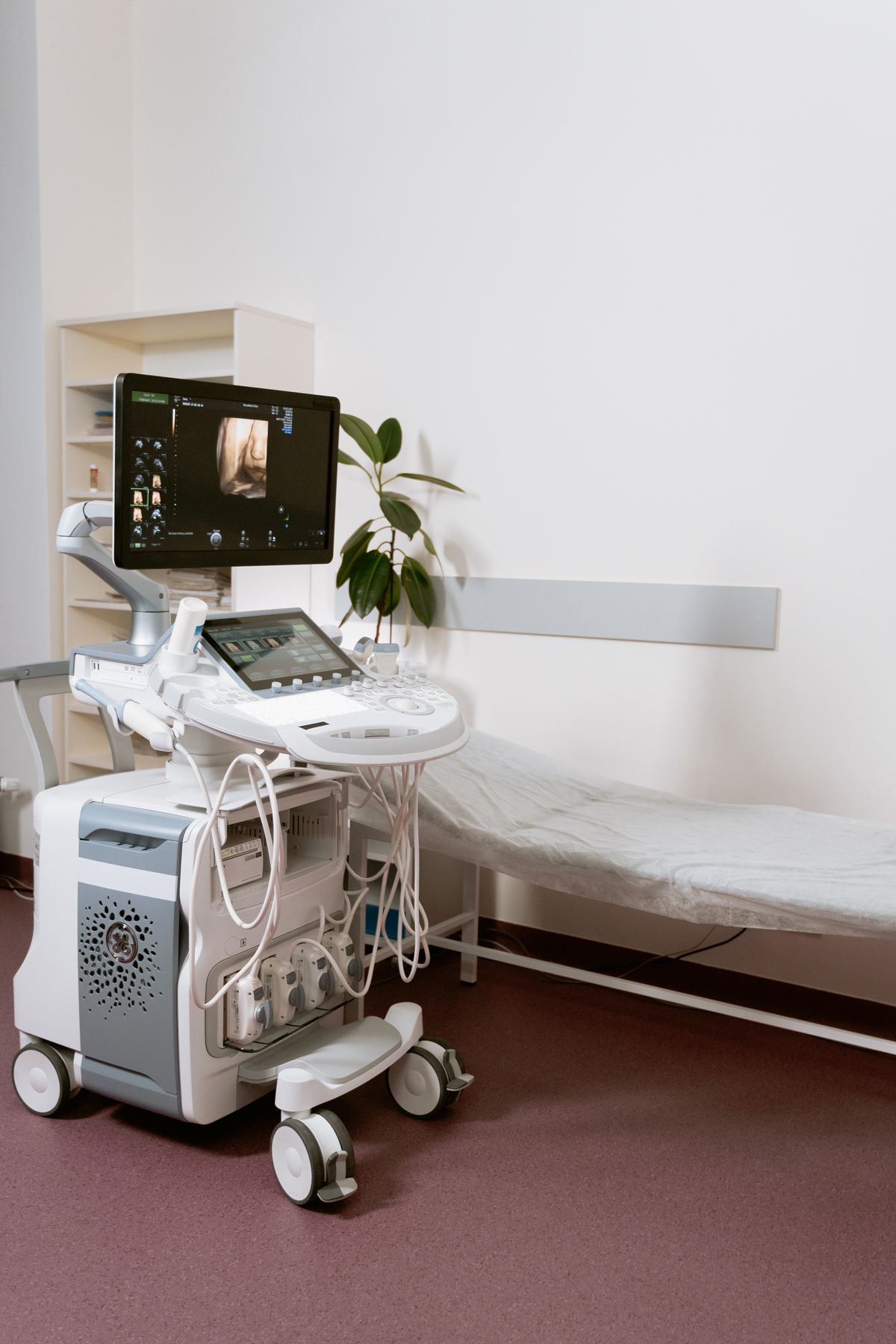
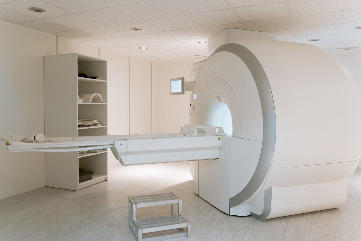
A Picture Archiving and Communication System (PACS) is a medical imaging technology used to store, retrieve, distribute, and present images from various modalities (X-ray, CT, MRI, ultrasound). PACS has revolutionized radiology by replacing traditional film-based imaging with digital workflows, enabling faster diagnosis and better patient care.
Core Components
Imaging Modalities
- X-ray, CT, MRI, ultrasound, nuclear medicine
- Mammography and fluoroscopy systems
- Each modality generates DICOM-format images
Acquisition Gateway
- Receives images from modalities
- Validates DICOM compliance
- Routes images to archive and workstations
Archive Server
- Long-term image storage (online, nearline, offline tiers)
- Database for image metadata and patient records
- Data compression and encryption
- Backup and disaster recovery
Workstations
- Diagnostic workstations for radiologists
- Review workstations for referring physicians
- Web-based viewers for remote access
- Mobile viewers for on-call physicians
Network Infrastructure
- High-speed network for image transfer
- Secure connections between modalities and archive
- Bandwidth management for large image files
DICOM Standard
DICOM (Digital Imaging and Communications in Medicine) is the international standard for medical images:
- Defines image format and metadata
- Specifies network communication protocols
- Includes patient demographics and study information
- Supports compression (lossless and lossy)
- Enables interoperability between vendors
PACS Workflow
- Image Acquisition: Modality generates DICOM images
- Image Routing: Gateway sends images to archive
- Image Storage: Archive stores images with metadata
- Image Distribution: Images sent to appropriate workstations
- Interpretation: Radiologist reads and reports
- Report Distribution: Report sent to referring physician
- Long-term Storage: Images retained per regulatory requirements
Integration with Other Systems
RIS (Radiology Information System)
- Manages patient scheduling and registration
- Tracks exam status and reporting
- Interfaces with PACS for worklist management
HIS (Hospital Information System)
- Patient demographics and clinical data
- Order entry for imaging studies
- Results distribution to electronic health records
EHR (Electronic Health Record)
- Embeds images and reports in patient records
- Enables physician access to imaging history
Benefits of PACS
- Faster diagnosis: Images available instantly
- Improved productivity: No film handling or storage
- Better collaboration: Remote access for consultations
- Cost savings: Eliminates film and chemical costs
- Enhanced patient safety: No lost films, better tracking
- Disaster recovery: Digital backup and redundancy
Implementation Challenges
- High initial investment in infrastructure
- Integration complexity with existing systems
- Staff training and workflow changes
- Data migration from film archives
- Cybersecurity and patient privacy (HIPAA compliance)
- Storage capacity planning for growing image volumes
Future Trends
- Cloud-based PACS: Reducing on-premise infrastructure
- AI integration: Computer-aided detection and diagnosis
- Enterprise imaging: Extending PACS beyond radiology
- Mobile access: Smartphone and tablet viewing
- 3D and advanced visualization: Volumetric rendering
Conclusion
PACS is a cornerstone of modern healthcare IT, enabling efficient medical imaging workflows that improve diagnostic speed, patient care, and operational efficiency. Understanding PACS architecture, DICOM standards, and integration requirements is essential for healthcare IT professionals and project managers implementing these systems.